
A Secondary 1 student in Punggol draws a perfect red blood cell and writes beside it, “This is an organ because it helps the body.” There is a hopeful instinct behind the mistake: the student knows that this tiny structure matters. But the answer also reveals a missing map. What makes a cell different from a tissue? How do tissues make an organ? Why does an organ need the rest of a system? Biology begins to make sense when a learner can move confidently between those levels.
Secondary 1 Punggol Biology tuition is most valuable when a child needs targeted help with specialised cells, cell structure and function, tissues, organs, organ systems, red blood cells and Lower Secondary Science. In mainstream Singapore secondary schools, these topics are usually taught within integrated Science rather than a separate compulsory Pure Biology subject. The learning aim is therefore to strengthen the correct G1, G2 or G3 curriculum foundation, distinguish a model cell from a real specialised cell, and develop explanations that transfer to unfamiliar questions. More technical vocabulary is useful only when the student understands what each level of organisation means.
The lovely thing about this topic is that it turns a living organism from one enormous mystery into a collection of smaller cooperating parts. A red blood cell has one job; cardiac muscle cells another. Organs combine different tissues, and organ systems coordinate functions that a single cell cannot perform alone. The first triumph is not remembering a diagram. It is being able to explain why cooperation is necessary.
Follow the four-year Punggol Biology series: Secondary 1: Specialised Cells and Systems · Secondary 2: Human Reproduction and Puberty · Secondary 3: Photosynthesis and Limiting Factors · Secondary 4: Mitosis, Meiosis and Inheritance
Why specialised cells can be the missing connection
A student may memorise that a typical animal cell has a cell membrane, cytoplasm and nucleus, then conclude that every cell in every animal looks exactly like the textbook sketch. The diagram is useful, but it is a model. Specialised cells differ in shape, structure and function. Some depart strikingly from the representative drawing.
That distinction becomes important when a school Science question presents an unfamiliar picture. A pupil might look at a long muscle fibre or a biconcave red blood cell and fail to recognise it as a cell. Another may correctly identify it but cannot relate its features to its job.
The best reason for tuition is a repeated difficulty in connecting structure to function or cells to higher levels of organisation. A targeted tutor can show the missing relationship, then ask the learner to apply it to a new cell type. The aim is independent thinking, not simply filling pages with labelled pictures.
If your child already does that confidently and is coping with school, extra lessons may not be needed. Enjoyable reading, school feedback and regular retrieval can be sufficient.
The MOE syllabus distinction every parent should know
The MOE G2/G3 Lower Secondary Science syllabus includes Model of Cells — the Basic Unit of Life. It requires students to recognise that cells in multicellular organisms are organised into tissues, organs and systems, while explaining division of labour at the cellular level is marked optional for G2. G2 pupils should not automatically be assessed against every additional G3 elaboration.
The MOE G1 Lower Secondary Science syllabus has a topic on Cells that explicitly includes the functions of bone cells, muscle cells and red blood cells, as well as their organisation into tissues, organs and systems. It uses a different level-appropriate route.
These are *lower-secondary stage* specifications. Schools determine their classroom order and assessment timetable. A responsible tutor checks the learner’s actual notes, teacher instructions, subject level and independent work before deciding what needs repair.
The hierarchy students need to reconstruct
A useful explanation begins with a clear scale:
Cells → tissues → organs → organ systems → organism.
A cell is a basic structural and functional unit of life. A tissue is a group of cells organised for a related function. An organ consists of different tissues contributing to particular functions. An organ system includes organs cooperating in a larger biological task. The organism is the living whole.
A muscle cell is not an organ. Muscle tissue is not an organ system. The heart is an organ containing different tissues; the circulatory system includes the heart and blood vessels, with blood and related components contributing to transport.
The tutor should not ask students merely to chant five nouns. Give a concrete example at each level and explain the relationship between adjacent levels. If the child can rebuild the sequence with an unfamiliar system, the terminology has become usable.
Worked question: why a red blood cell is not an organ
Question: A pupil says, “A red blood cell carries oxygen, so it is an organ.” Explain why the reasoning is incorrect.
Incomplete answer: “Red blood cells are smaller than organs.”
Stronger answer: “A red blood cell is a specialised cell. An organ is made of multiple tissues organised to perform functions. Although a red blood cell contributes to oxygen transport, that function does not make an individual cell an organ.”
The better answer explains a category difference, not merely a difference in size. A large cell is still a cell; a tiny organ is still an organ. Function matters, but biological organisation matters too.
Next ask the learner to classify a piece of muscle tissue, a kidney and the urinary system. A correct changed answer demonstrates transfer.
What makes a specialised cell specialised?
A specialised cell possesses structural features suited to its particular role. Some cells are elongated, some have a large surface area, and some possess different numbers or arrangements of structures. Their form contributes to how they perform specific functions.
A human red blood cell, for instance, has a biconcave shape and contains haemoglobin for oxygen transport. Mature human red blood cells lack a nucleus, showing why a model animal cell with a prominent nucleus cannot represent every specialised animal cell.
A muscle cell is adapted for contraction, contributing to movement or the function of an organ such as the heart. The different types of muscle have distinct biological properties, but the first lower-secondary lesson is about relating structure and task.
A bone cell contributes to the maintenance and function of bone tissue. Pupils do not need a university-level account of every bone-cell type unless extension is expressly appropriate.
Teach the feature–function connection and keep the details proportionate to the school’s syllabus.
Typical cell diagrams are useful because they simplify
A textbook illustration is not trying to photograph every kind of animal cell. It helps learners recognise a representative nucleus, cytoplasm and cell membrane and discuss their main roles. In a typical plant-cell model, features such as a wall and a large vacuole may also be shown.
A pupil who sees a mature red blood cell and says, “It cannot be an animal cell because the nucleus is missing,” has mistaken the model for an absolute rule. The tutor can explain what makes the cell an animal cell and why specialisation can involve a structure differing from the typical diagram.
This is a deeper scientific skill: models explain important patterns but have boundaries. A good model helps us think, yet it does not show every real exception.
That habit will later help with food webs, particulate models, graphs and even mathematical representations. Children benefit from learning when a simplified picture is informative and when it is incomplete.
Red blood cells: three reasons their form matters
In the familiar human biology example, the red blood cell’s biconcave shape creates a favourable surface-area-to-volume relationship and a short diffusion pathway for oxygen. Its flexible form helps it move through narrow capillaries. Haemoglobin provides a mechanism for binding and transporting oxygen.
A student should avoid a tempting overstatement: “Red blood cells contain the oxygen that they make.” Red blood cells do not manufacture the oxygen; they transport oxygen acquired through the respiratory system.
Similarly, the loss of a nucleus in a mature human red blood cell is a specialised characteristic, not a rule for every animal cell. Its physiological features suit transport, although real biological function is more complex than any one-line mnemonic.
If the student’s present syllabus requires only a general function, begin there. Additional explanation should strengthen comprehension rather than become a new list of unnecessary memorisation.
Muscle cells: how function is more than a label
The word muscle appears everywhere in daily speech: leg muscles, heart muscle, movement, exercise. In Science, the important function is the ability of muscle tissue to contract and contribute to movement or other bodily activity.
A tutor might ask why the wall of the heart cannot be made entirely from ordinary surface-lining cells. The intended answer is not a complete anatomy lecture. It is that different cells have structural and functional properties needed for different tasks.
Now ask the student to connect this back to the biological hierarchy. A muscle cell contributes to muscle tissue, which may be part of an organ, which may contribute to an organ system.
This prevents the pupil from treating the hierarchy as a ladder of unrelated words. It is a nested arrangement of working biological structures.
Bone cells: the quiet workers in a living tissue
Children often think of bones as inert sticks holding up the body. Bone is living tissue with cells and a specialised extracellular matrix. Different bone-associated cells help maintain, form or remodel the tissue, though detailed classification is not necessary for every lower-secondary learner.
The broad educational point is that bone cells contribute to the structure and maintenance of bone tissue, which supports and protects the body and works with other structures in movement.
Ask the child whether bone is a cell or an organ. The entire bone is an organ composed of several tissues; bone tissue includes cells and matrix. This may feel like an awkward distinction initially, which is why it is useful.
A clear explanation helps students recognise the same organisational principles in organs beyond the skeleton.
The heart: one organ, more than one kind of tissue
The heart pumps blood, but it is not a giant cell. It contains different tissues working together, including cardiac muscle tissue involved in contraction and other tissue types contributing to structure and function.
A student may know that the heart beats and blood moves, yet miss the relationship between the cellular properties of muscle and the function of the organ. Linking the two creates a new explanatory path.
From there, move to the circulatory system. It includes the heart and blood vessels, with blood acting as an important transport medium. The system supports distribution of substances that cells need.
The learner should be able to describe a path from specialised cell → tissue → organ → system function without claiming that any one cell performs every task in the whole organism.
Why organ systems must cooperate
A circulatory system cannot supply oxygen that never entered the body. Gas exchange through the respiratory system helps oxygen reach blood, and circulatory transport can carry it towards tissues. Digested nutrients can enter transport pathways after relevant digestive processes.
The body therefore needs different systems with complementary roles. This is a good opportunity to show why knowing one textbook chapter is not always enough for a new question.
An excellent prompt is: “How do the lungs and circulatory system cooperate to help an active muscle cell?” The student should explain air-to-blood gas exchange, transport and cellular use without saying the lungs directly push oxygen into every cell.
At Secondary 1, the required depth should follow school Science content. The point is cooperation, not early examination drilling from an advanced Biology paper.
Specialisation is an example of division of labour
Division of labour means different components perform different roles that contribute to a wider function. Biology contains many examples of specialised cells, but students should remember that the formal significance of division of labour is an optional G2 outcome in the MOE G2/G3 lower-secondary syllabus.
A tutor can illustrate the idea through a non-biological analogy, such as a school organising a performance. Different people handle sound, lighting and stage management. No single person needs to perform every job, and cooperation enables the event.
The analogy must then return to biology. Cells are not miniature conscious workers who choose their tasks. Their structures, gene expression and developmental pathways underlie specialisation.
A good analogy opens a door; a clear biological explanation stops it from becoming a misconception.
How a microscope image can create confusion
Prepared images of cells can be difficult to interpret. Some specialised cells look very different from textbook models, and some structures are too small to be resolved clearly by an ordinary school light microscope.
A student may be tempted to add labels from memory to features not actually visible. Encourage a distinction between observed feature, reasonable identification and structure known from the model.
For example, a red blood cell image may show its general outline without directly demonstrating haemoglobin molecules. The learner may know about haemoglobin from established Biology, but should not say that an individual molecule was visibly identified in that ordinary image.
This is a simple introduction to scientific honesty. It also improves data-based answers: identify what the evidence shows before claiming an explanation.
One diagnostic exercise across four levels
Present a short set of cards: “red blood cell,” “muscle tissue,” “heart,” “circulatory system,” “bone,” “bone cell,” “lungs” and “respiratory system.”
Ask the learner to classify each at the appropriate organisational level. Then choose one group and explain how the levels are connected. Avoid mixing the biological roles: a red blood cell is not a kind of heart muscle cell, even though both contribute to circulation.
The cards are only the opening. Next present a new item, such as a leaf or a plant transport tissue. Can the student transfer the classification logic to plant biology?
A pupil who can explain the new example without hints is displaying more than rote memory.
A structured-answer clinic for Secondary 1 Science
Question: Explain why different kinds of specialised cells are useful in a multicellular organism.
Weak answer: “There are lots of cells, so they all have jobs.”
Stronger answer: “Specialised cells have features suited to different functions. In a multicellular organism, different cells can contribute to tissues, organs and systems that perform complementary tasks, allowing the organism to carry out functions that an individual cell alone could not perform.”
The stronger response uses the key idea of different functions and their coordination. For G2 learners, a tutor should be clear if this detailed division-of-labour explanation is being taught as enrichment rather than as a required outcome.
An independent retest could ask about a plant root or an organ in an unfamiliar animal. A copied sentence is not the destination.
Four common mistakes and what to teach instead
“Every cell looks like the model.” Teach what a model represents and why specialised cells can differ.
“Anything that performs a job is an organ.” Teach the organisational definition of an organ as a structure comprising tissues.
“A bigger structure is automatically a system.” Teach relationships: organs working together form organ systems, not merely large objects.
“Cells in an organ all do the same thing.” Discuss how different tissue types contribute to the organ’s function, using appropriate examples.
Each of these errors can be repaired through one clear contrast followed by an unfamiliar question. The number of pages completed matters less than the learner’s ability to correct the original misconception independently.
What a small-group lesson can add
Imagine three pupils looking at the same cell-to-system diagram. One student confuses tissue with organ. A second thinks all animal cells contain a nucleus. A third knows the definitions but cannot connect muscle tissue to heart function.
A three-student format can allow the tutor to observe each response before explaining. The pupils then receive different corrective tasks and individual follow-up questions, rather than silently copying one answer.
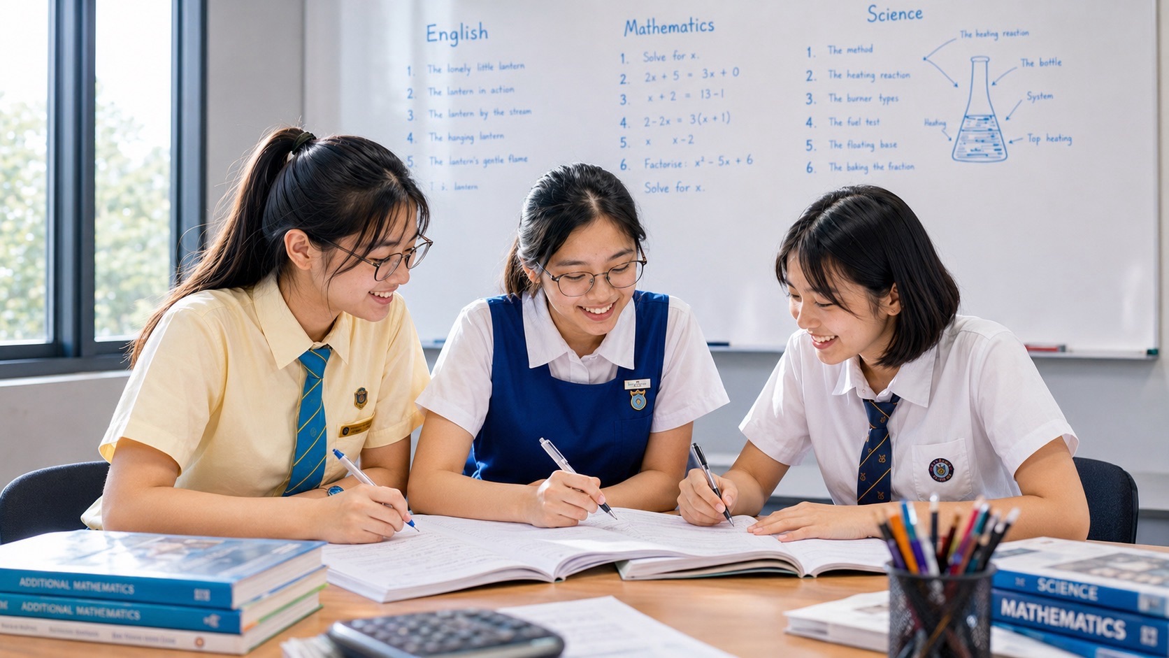
The immutable eduKateSG small-group tutorial reference illustrates this diagnosis–correction–transfer principle in Mathematics. The method can inform Science tuition, but that reference does not confirm a specific Biology class, timetable or available seat in Punggol.
Parents can ask about the actual programme at the eduKatePunggol tuition hub. A useful class should be able to show independent improvement, not merely offer the label “small group.”
A six-week foundation plan
Week 1 — Locate the first gap. Examine the child’s school-level cell and organisation questions. Test distinctions among cell, tissue, organ and system.
Week 2 — Revisit representative cells. Compare a typical animal or plant cell with a real specialised-cell example, at the actual G-level scope.
Week 3 — Connect features with functions. Use red blood, muscle and bone examples where appropriate to the syllabus.
Week 4 — Organise into systems. Build a complete example from cells through a tissue and organ to an organ-system function.
Week 5 — Change the context. Introduce an unfamiliar plant or animal example and require accurate classification and explanation.
Week 6 — Retest independently. Compare changed questions with the original baseline. Continue only where a specific need remains.
This is an illustrative route, not an MOE-imposed sequence or a promise that such a course is currently scheduled. School topics may be taught in a different order.
Parents’ questions about specialised cells and tuition
Is specialised cell structure part of Secondary 1 Biology?
It is relevant to lower-secondary Science and its cell models. The exact depth depends on G1, G2 or G3 scope and the school’s sequence. In G2/G3, explaining the significance of division of labour at cellular level is specifically optional for G2.
Is the heart a cell, tissue, organ or system?
The heart is an organ composed of different tissues. It works within the circulatory system.
Are red blood cells typical animal cells?
They are specialised animal cells. Mature human red blood cells lack nuclei, so they are not identical to the typical textbook animal-cell model.
Do G1 pupils study specialised bone and muscle cells?
The MOE G1 lower-secondary Science cells topic explicitly includes the functions of bone cells, muscle cells and red blood cells.
Does a bigger cell become a tissue?
No. A tissue is an organised grouping of cells carrying out related functions. Size alone does not change the biological category.
Should the child memorise advanced organ histology now?
Only if it aligns with the child’s syllabus or genuinely useful extension. Correct understanding of the current levels of organisation comes first.
Can a student learn this without private tuition?
Yes. Current school notes, teacher feedback and changed independent practice questions can be sufficient. Tuition is most appropriate when a specific misconception persists.
What evidence shows tuition has helped?
The learner should accurately explain unfamiliar specialised-cell examples, distinguish organisational levels and show improved independent answers after a delay.
The next reading route
For foundational context, read Secondary 1 Microscope Skills and Cell Diagrams and Secondary 1 Lower Secondary Science and Cell Biology. To see how body systems become connected, continue into Secondary 2 Human Circulatory System and Blood Vessels and Secondary 2 Human Digestive System and Nutrient Absorption.
The MOE G2/G3 Lower Secondary Science syllabus and MOE G1 syllabus remain the official scope references.
The moment a diagram becomes a living system
The red blood cell in the opening story was not an organ, but the student’s instinct that it was important was correct. Biology tuition can preserve that curiosity while giving it a reliable structure: cells have functions, tissues bring cells together, organs coordinate tissues, and systems bring organs into cooperation.
When a Secondary 1 learner can explain that structure in a new example, they are no longer simply memorising Science. They are beginning to understand how living systems work—and why a good explanation lasts much longer than a correctly coloured diagram.


